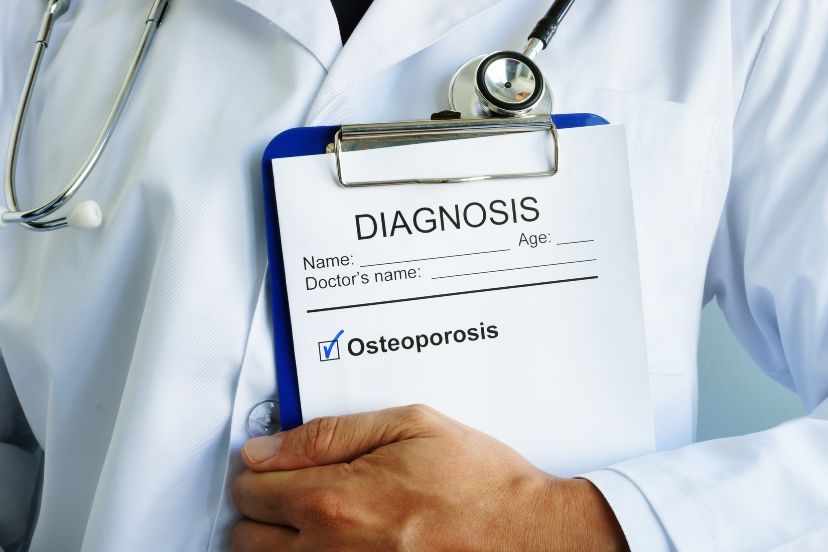
Osteoporosis Diagnosis: Understanding the Process
Introduction
Welcome to my comprehensive guide on how osteoporosis, a condition characterized by weakened bones and increased fracture risk, is diagnosed. As someone passionate about promoting bone health, I understand the importance of an early osteoporosis diagnosis and timely intervention. In this article, we will delve into the various diagnostic methods employed by healthcare professionals to identify osteoporosis. From bone density tests to medical history assessments, physical exams, and advanced imaging techniques, we will explore each approach to provide you with a holistic understanding of the diagnostic process. By the end, you will be equipped with the knowledge necessary to actively engage in your bone health and advocate for early detection.
The Role of Bone Density Tests
Bone density tests, also known as dual-energy X-ray absorptiometry (DXA) scans, play a vital role in diagnosing osteoporosis. These tests measure the mineral content and density of your bones, providing valuable information about their strength and susceptibility to fractures. DXA scans are typically conducted on the hip and spine, as these are common sites of osteoporotic fractures. The results are compared to the average bone density of young adults, generating a T-score that indicates your bone health status.
Medical History Assessment
A thorough evaluation of your medical history is crucial in diagnosing osteoporosis. Your healthcare provider will ask about your personal and family medical history, including any previous fractures, hormonal disorders, and lifestyle factors such as smoking, excessive alcohol consumption, and sedentary behavior. Understanding your risk factors enables healthcare professionals to tailor diagnostic and treatment approaches to your specific needs.
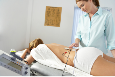
Comprehensive Physical Exams
Physical exams are an essential component of the diagnostic process for osteoporosis. During the exam, your healthcare provider will assess your height, posture, and overall musculoskeletal health. They may look for signs of vertebral compression fractures, such as a loss of height or curvature of the spine. Additionally, they may evaluate your balance and muscle strength, which are essential for maintaining bone health and preventing falls.
Laboratory Tests
Laboratory tests can help identify underlying causes of bone loss and assess the overall health of your bones. Blood tests are commonly performed to measure levels of calcium, vitamin D, and other minerals involved in bone metabolism. These tests can also determine if there are any underlying medical conditions contributing to bone loss, such as hyperthyroidism or kidney disease.
Imaging Techniques for Bone Loss Detection
In some cases, additional imaging techniques may be employed to detect osteoporosis and evaluate the severity of bone loss. These techniques include:
X-Rays:
X-rays can reveal fractures, bone deformities, or vertebral collapses. However, X-rays alone are not sufficient to diagnose osteoporosis. They are typically used in conjunction with other diagnostic methods.
Quantitative Ultrasound (QUS):
QUS is a non-invasive technique that measures bone density using sound waves. It is often used to assess the heel bone and predict fracture risk.
Magnetic Resonance Imaging (MRI):
MRI scans can provide detailed images of the bones, helping detect fractures, infections, or tumors that may contribute to bone loss. While not commonly used for routine osteoporosis diagnosis, they are valuable in specific cases.
Computed Tomography (CT) Scan:
CT scans can assess bone density and detect fractures with high precision. However, due to the increased radiation exposure compared to DXA scans, they are usually reserved for specific situations.
Conclusion
Early osteoporosis diagnosis is vital for effective management and prevention of fractures. Through a combination of bone density tests, medical history assessments, physical exams, laboratory tests, and imaging techniques, healthcare professionals can accurately diagnose osteoporosis and tailor treatment plans to individual needs. Remember, understanding the diagnostic process empowers you to take proactive steps toward maintaining healthy bones and reducing fracture risk. Advocate for regular screenings and discuss any concerns or risk factors with your healthcare provider.
FAQs (Frequently Asked Questions)
Q. Are bone density tests painful?
A: Bone density tests are painless and non-invasive. They involve lying on a padded table while a machine scans your bones with low levels of radiation.
Q. Can osteoporosis be diagnosed without a bone density test?
A: While bone density tests are the gold standard for diagnosing osteoporosis, other factors such as medical history, physical exams, and laboratory tests can contribute to an accurate diagnosis.
Q. How often should I undergo bone density testing?
A: The frequency of bone density testing depends on various factors, including your age, sex, and overall fracture risk. Generally, postmenopausal women may be recommended to have a DXA scan every 2-5 years.
Q. Can I prevent osteoporosis even if I’m diagnosed with the condition?
A: Although osteoporosis cannot be reversed, its progression can be slowed down, and the risk of fractures can be reduced through a combination of lifestyle changes, including a healthy diet, regular exercise, and appropriate medical interventions.
Q. Can men get osteoporosis?
A: Absolutely! Although osteoporosis is more common in women, men can also develop the condition. It is important for both men and women to prioritize bone health and discuss any concerns with their healthcare providers.